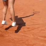
The “calf” muscle is composed of the soleus, gastrocnemius and plantaris muscles. Together they form the achilles tendon and insert into the calcaneus. Contraction of these muscles results in ankle plantar flexion. In the middle aged and intermittently active tennis player, the soft tissues of the leg often lack the elasticity and flexibility required to safely perform the tasks demanded during play. As a result when the individual sprints toward a ball they may suddenly feel a “pull” or as though “shot in the leg” in the back leg and immediately be unable to weight bear without significant pain.
They often present for clinical evaluation limping or using crutches and with a region of ecchymosis and mild swelling over the posterior leg. Pain, swelling or ecchymosis near the calcaneus should prompt a careful evaluation of the achilles tendon for possible tendon rupture (inspect for defect in achilles bulk, ecchymosis over distal leg, focal swelling over achilles, + Thompson test, Ultrasound evidence of tear etc.). If the patient has swelling of the entire leg then alternative diagnosis such as a DVT (deep venous thrombosis) must be considered (+Homan’s test, personal or family history of DVT’s, use of birth control, recent prolonged immobility etc.). The patient with “tennis leg” should present with mild to significant pain with weight bearing, worst with “toe-off” during stride or active ankle plantar flexion. The patient will also have tenderness most commonly over the medial proximal/mid-calf region with an otherwise normal neuro-vascular exam of the leg and with normal X-rays of the leg, negative heel jar and percussion testing over bony elements (tibia/fibular head). If the clinical history/exam are consistent with “tennis leg” and alternative diagnosis have been ruled out then treatment should be started.
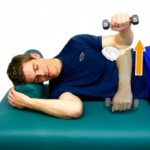
Treatment includes protected weight bearing during the acute phase with crutches or a cane and limitation of ankle dorsiflexion with a boot or soft tissue splint. Ankle range of motion is limited for 1-2 weeks to allow rapid “scarring” of the torn region and to reduce the risk of a chronic seroma. We support aggressive icing for pain management and encourage avoidance of NSAIDs since the acute “inflammatory” cascade of platelets etc promote rapid healing. If minimal pain on exam, normal or near normal gait, no focal defects in the muscle and a normal ultrasound exam, then the patient may begin an isometric followed by eccentric calf strengthening program. If significant pain and debility on initial exam the paint should be aggressively rested as described above for 1-2 weeks and then re-examined in the clinic. Once the patient has minimal pain then a progressive rehabilitation program should be started.
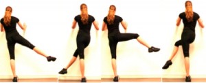
Prevention is key and includes a five-ten minute cardiovascular warm up prior to play including a dynamic stretching program and both plyometric training and focused strengthening of the calf as featured on our site.